EXPERT SHARK PARASITOLOGIST VISITS DYER ISLAND CONSERVATION TRUST / MARINE DYNAMICS
June 04, 2010 by dyertrust
World renowned shark parasitologist, Professor Susan Dippenaar, from theUniversity of Limpopowas invited to visit Dyer Island Conservation Trust /Marine Dynamicsto advise our researcherHennie Ottoon his study of parasites on the great white shark (Carcharodon carcharias). Prof. Dippenaar specialises in siphonostomatoids, a tongue-twisting name for a small part of a larger group of animals commonly known as copepods.
Copepods are the most common and abundant animal group found in the marine and freshwater environments. They form such a large part of the zooplankton that it is reckoned that copepods have the largest biomass of any animal group on earth. Many of the 12,000 or so species are planktonic (drift in sea water) and are an important part of marine food chains. They are a major source of food for a variety of animals like seabirds, whales and most fish species including the largest of all known living fishes, the whale shark. Apart from their nutritional value, copepods are also instrumental in the natural carbon cycle. They feed on photosynthetic algae at night, allowing faeces, respiration, moulted exoskeletons and dead bodies to sink to the ocean floor. Deep sea carbon storage forms an integral part of natures way to avert global warming.
To date several species of copepods have been described from great white sharks, (Carcharodon carcharias). These include:
Alebion carchariae, Anthosoma crassum, Dinemoura latifolia, Dinemoura producta, Echthrogaleus coleoptratus, Echthrogaleus denticulatus, Nemesis lamna, Nesippus orientalis, Pandarus bicolour, Pandarus cranchii, Pandarus satyrus, Pandarus smithii and Achtheinus oblongus (Dippenaar 2005 & Hewitt 1979).
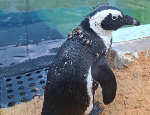
The host shark provides a matrix of potential habitats for parasitic organisms. Parasites have been found on the epidermal surface, gill arches and gill lamellae, inside nasal passages and the mouth amongst the teeth. It is hypothesised that certain siphonostomatoids move freely around on the skin on infected great white sharks and feed on mucus and the epidermal surface. High quality, high resolution images of free-swimming great white sharks carrying obvious infections of siphonostomatoids are taken as they interact with our cage diving vessel,Shark Fever.
Traditional field-based siphonostomatoids – great white shark associative studies requires incapacitated host specimens, many of which are captured in nets/deceased.
The video clip indicates obvious infections of what appears to be Pandarus spp. attached to the pectoral fins, head, gill area and the tail of a great white shark. This is an alternative non-invasive study method of visual identification and distribution obvious parasitic infections through digital imagery. The patterns in the dynamics of infection are recorded in space and time. Our study will not only focus on the great white shark, but also include multiple species of commercially exploited sharks like blue sharks, mako, bronze whaler, soupfin and seven gill cow sharks.

From left to right: Bea Jordaan (research technician-University of Limpopo); Hennie Otto (DICT/Marine Dynamics); Prof. Susan Dippenaar (University of Limpopo)

Kroyeria carchariaeglauci found on the gill filaments of a blue shark in Gansbaai
